Causes

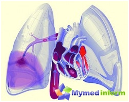
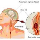
In order for the human body normally, it should continuously get the required amount of oxygen. Oxygen is supplied to the body usually through the gas exchange process in the lungs. In order for oxygen to flow from the lungs to the authorities, there is blood supply. With the thromboembolism of the lungs at the site of the lesion, the normal blood supply does not occur, which leads to a violation of gas exchange. Light, and, therefore, other organs get less oxygen. Blood, which passes through the vessels because of this, is much worse than saturated with oxygen. Internal organs suffer from oxygen deficiency. As a result, arterial pressure can sharply decrease, which in turn leads to Myocardial infarction and atelectasia in the lungs. Thromboembolism of the lungs most often arises due to thromboms that were formed in the veins of the legs. To form thrombus, it is necessary that the blood flow is slow enough, blood clotting was increased and the walls of the vessels were damaged.
Normal blood flow, often slowed down due to heart failure. In addition, it can be disturbed in people who are long enough in one position. For example, if a person is forced to observe the bed. Most often it occurs in patients who have damage to the spinal cord. Quite rare, but it is still possible to thromboembolism even in healthy people who have long been in the same position. For example, with a long flight by plane. As for damage to the walls of the veins, this is often happening in intravenous injections, inflammatory diseases and injuries. Increased blood clotting is most often evolving due to violations in a blood coagulation system. In addition, the reason for this may become AIDS and reception of contraceptives.
In addition to the specified risk factors, the peace and the elderly and senile age, malignant education, surgical operations, phlebeurysm, Various injuries, pregnancy and childbirth, obesity And some diseases.
Symptoms

In the presence of characteristic symptoms of the disease, some studies are held. In particular, with the help of the chest x-ray, it is possible to detect small changes in the structure of blood vessels after embolism. In addition, this procedure allows you to identify a light infarction. However, when these symptoms are detected, it is impossible to put an accurate diagnosis. Since to confirm the presence of changes in vessels, an electrocardiogram is carried out, perfusion scintigraphy and other diagnostic methods.
In order to carry out perfusion scintigraphy of the lungs, which is also quite useful in the diagnosis of pulmonary thromboembolism, it is necessary to introduce a small amount of radionucleic substance to Vienna. The substance along with blood enters the easy, which makes it possible to assess the blood supply to the lungs. If, when conducting this procedure, areas were revealed, where there is no normal blood supply, then in the picture they will be dark. This is due to the fact that radionucleic particles do not fall into the zones with poor blood supply. If there are no results of such areas, it is believed that a person has no significant blockage of blood vessels. However, the identified signs of blockage of vessels can be associated with other violations in the body, and not only indicate the presence of pulmonary artery thromboembolism.
Treatment

For the treatment of thromboembolism of the patient, they are usually directed to the hospital. This is necessary because the timely treatment is largely depends on how the disease will develop in the future and is it possible to survive the patient.
In the treatment, conservative therapy techniques, surgical therapy and, if necessary, resuscitation activities can be used. Almost in all cases in the treatment of pulmonary artery embolism, such a drug as heparin or its analogues is used. In conservative treatment, fibrinolytic drugs are used. As for surgical treatment, it is usually prescribed only with critical blood flow disorders by vessels, with severe cardiovary pulmonary failure, with extensive lesions of the pulmonary artery, in the absence of time for thrombolysis. This kind of treatment should be carried out only by experienced and prepared surgeons.
To prevent the formation of thromboms in the legs of the tibia after operation, such an opposing agent is used as heparin. Before the operation and within a week after the operation, the patient is entered by small doses of this drug under the skin. True, heparin is usually appointed only by patients who have a high risk of thrombosis, patients with heart failure, suffering from obesity, with chronic lung diseases and patients who had cases of thrombosis before. This is due to the fact that this drug can cause bleeding and slow down healing. With a high risk of the development of the pulmonary embolism, small doses of heparin are sometimes prescribed, and even if the operation is not expected. It is worth noting that approximately half of all patients with pulmonary artery embolism that have not received normal treatment, there is a high risk of re-development of embolism.


