What is the Doppler effect?
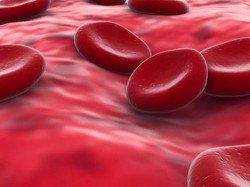
The basis of this body examination is the so-called Doppler effect. It was described in the middle of the nineteenth century Christian Doppler (Austrian physicist). The essence is as follows: when the receiver or the source of the waves moves, changes and the frequency of oscillations occurs. Accounting for these changes and is carried out during dopplerography. The sensor captures reflected waves, and the computer on the basis of the data obtained displays the picture (black and white or color). Modern devices are able to simultaneously analyze information and still structures (objects), as it happens when ultrasound, and in the end they give out the overall consolidated, meaningful picture for the doctor. A specialist using a certain hardware can see and assess the presence of pathological processes in vessels. Investment draws attention to the adjacent tissues. Color Card Flood allows you to determine any diseases of the vessels. The big plus of dopplerography is that with its help you can diagnose diseases even until the moment when you know about yourself clinical symptoms. It is possible to track the path of blood movement along the vascular bed, in parallel detecting disorders (if present). It can be understood that there are irregularities in vessels, their narrowing. You can even determine the speed of movement of the muscular walls and the state of various heart valves. Moreover, in contrast to angiography, dopplerography is harmless to the patient, contrast agents are not introduced into the bloodstream. The method is actively used in obstetric practice, the doctors have the opportunity to measure the blood flow rate in the vessels of the umbilical umbilical fetus and the uterine arteries. It is possible to register the movements of the heart of the fetus. And simply ultrasound scanning allows you to make only a visual assessment and measure the dimensions of the uterus, spindle water, placenta, the most developing fetus and its parts and organs.
Testimony to the survey
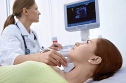
Cabinet, where you are conducting dopplerography, very similar to the one where they are engaged in ultrasound research. The duration of the procedure is usually 30 minutes. No diagnostic substances in the body are not required. Do not be afraid of pain and unpleasant sensations - they will not. In most cases, no preparations from the patient required. But there are devices equipped with special instructions. Sometimes the patient may ask not to dinner late a day preceding the survey. The obstacle for high-quality surveys may become cavity filled with accumulated gases (it happens in the lungs or intestines). To avoid such interference to the patient can recommend to take on the eve of the procedure (in the evening and morning) drugs, «Gassing» gases such as Sab Simplex, Espumizan. Patient first laid on the couch (position on the back). Under your head there will be a pad. The examination area will be treated with a gel that optimizes the contact between the sensor and the skin (it is used and when conducting an ultrasound). Then the sensor will be tightly pressed to the body, and the specialist's hand slowly moves (shifts) it in the same area to be examined. You will only sense some sensor pressure on skin. Sometimes you will hear a light whistled from the instruments. It means imitation of blood movement inside vessels. No restrictions of life after the procedure. You can do our usual affairs and responsibilities. No complications have been identified after such procedures.
What are the dopplers?
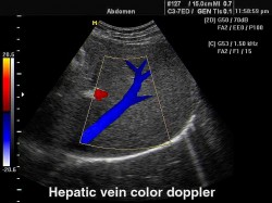
Color - this device allows you to identify blood violations by vessels (veins, arteries), examine the heart. On echoogram the type of blood flow is indicated by color. Red - the path of blood to the device, and the opposite direction is shown in blue. Other Procedure Name - Color Doppler Mapping.
Energy - With it, it is possible to make an assessment of the state blood circulation, when the speed of blood is small. Effective when examining ovaries, scrotics, thyroid, liver, kidneys. The method allows you to distinguish the cyst from the tumor. There are no blood vessels in the cyst, so it will not give orange glow.
Tissue - This doppler is useful in determining the activities of the heart muscle. Tissue research such a method helps to detect:
- Ischemia (Moreover, you can even determine whether the tissues of the heart muscle are viable);
- Dysfunction diastoles;
- cardiomyopathy;
- the presence of additional ducts;
- Systemic diseases of the heart;
- Returning the organ after transplantation.

If you need to determine the contractile ability of the heart, then such a survey is combined with a pulsed doppler.
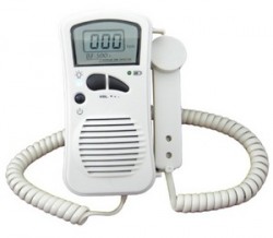
Analyzer - Before you start work, a specialist introduces the necessary parameters to this device for research. It is connected to a computer. You can control the device by resorting to the touchscreen display. After the examination, the data can be read on the printout, if necessary, they retain them to a digital removable device.
Fetal - a new-fashioned device that allows at least a round day, starting with the twelfth week of pregnancy, listen to how the fruit moves in the uterus, how his heart beats. Now many such devices are released by manufacturers. The developers assure that waves emanating from the devigor, are very weak, so do not harm or the future child nor his mom. The weight of the instrument does not exceed 150 grams, he has small dimensions. However, doctors are wary of such «Toys». They categorically do not recommend the device data to apply in the first 12 weeks of pregnancy. Even when the term passes during this period, often resort to the device undesirable. There are scientists who claim that the Doppler may harm. They believe that a strong ultrasonic wave can still disrupt the flow of important cell processes.
Hundreds of thousands of people are sent annually on the procedure of Doppler Studies.
Doppler software helps the doctors of different specializations in a timely manner and correctly diagnose and quickly begin treatment of their patients.