This article is addressed to all women, regardless of their age and health status. The reason for her writing is far from occasion - in our country in the last decade there is an increase in the incidence of breast cancer almost one and a half times. Now this diagnosis is made annually 50,000 women. And mammology beat alarm - mammary cancer Gradually, young people - now it can be found in women who have not reached 40 and even 30 years!
Is our mentality or something else, but the fact remains a fact: Russian women are still frivolous about their health, neglected even those in small preventive capabilities that the state provided them: the passage of mammography and dispensarization within the framework of free national programs «Health».
And nowhere now it is not a secret that almost everything in a timely identified (at the I-II stages of the development of the process) and treated malignant oncological diseases in 95% of cases end in full recovery. It also applies to breast cancer, the timely treatment of which allows to avoid coarse cosmetic defects and keep a woman an attractive appearance.
Some more statistics
As known, Mastopathy - Very common breast disease, which can cause cancer and is diagnosed with 45-60% of fertile women (25-55 years), as well as 90% of women with gynecological diseases. The detection of breast cancer with mammography even with small (less than 1 cm in diameter), non-fallen tumors is 95%. It is to identify the disease at this stage and it is necessary to undergo prophylactic inspections by mammologist.
Symptoms of dairy glasses
The main thing is that you need to learn - to avoid the serious effects of diseases of the dairy glands. But for this you should not wait when something sits. Attend a mammologist in preventive purposes is necessary at least once a year. The urgent appeal to the specialist require the following symptoms:
- The appearance of the selection from the nipple outside the breastfeeding period;
- The emergence of pain in the chest and feelings of discomfort, cutting, regardless of the phase of the menstrual cycle;
- Availability of rash, hyperemia, appearance «Lemon crust» or ulcers on the skin of one or both of the mammary glands;
- the appearance of nodes, seals in the tissues of one or both of the mammary glands, as well as under the skin or in the axillary depressions;
- the appearance of asymmetry and / or deformation of breast contours, its swelling;
- change of shape, contours or skin of the nipples, the appearance of their retractions or ulcerations;
- The appearance of enlarged (palpable) lymph nodes in the above and subclavian regions.
Factors contributing to the development of diseases
Unfortunately, our entire modern life with a worsening environment, an uncontrolled medication, infinite stress creates a favorable background for the development of diseases. Risk factors abound, the following can be attributed:
- Stresses, long-term psychological loads, lacking, insomnia has a negative impact on the hormonal state of the woman and its body immunity,
- Abortion and chronic inflammatory diseases of the female genital sphere (mastitis, salpingooforites, adnexites, endometriosis and others). In women who have undergone more than three abortions throughout the life, the risk of mastopathy development increases by 3-4 times;
- violations of the hormonal status of the female organism, independent and uncontrolled reception, as well as the alternation of hormonal tableted contraceptives;
- hereditary predisposition;
- diseases of the digestive bodies that violate the synthesis of steroid hormones (hepatitis, cholangitis, cholecystitis, colitis and others), as well as violating the absorption of all necessary substances to develop, disorders the synthesis and absorption of vitamins, irreplaceable amino acids;
- Endocrine diseases and conditions accompanied Violation of metabolism (adrenal diseases, hypo-or hyperfunction of the thyroid gland, tumors of the internal secretion glands, hypertonic disease, diabetes, metabolic syndrome and t.D.);
- defective and poor Vitamins and microelements powered with frequent use of fast food products, as well as fried and smoked dishes, food intake, poor fiber (cereals, vegetable food and t.D.);
- Harmful habits leading to body intoxication (smoking, alcohol, long-term drug intake), as well as the passion for solarium, professional harmfulness.
Self-examination of the mammary glands

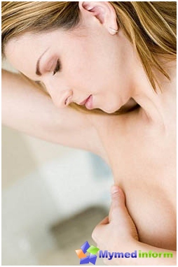
Every woman knows this. First of all, this method can be called a woman's responsibility for their own health. Self-examination of the mammary glands is a direct examination of the woman itself of its mammary glands, held every month, starting from 16-18 years during the first 2 weeks of the menstrual cycle. It includes inspection and consistent palpation of the glands for early detection of nodes and other changes in them. According to WHO statistics, it lowers mortality from malignant lactic gland neoplasms up to 20%.
Self-examination technique. No special skills required for this. Self-examination is carried out by the mirror, as well as while taking the shower and lying:
- To do this, undressing to the belt, standing with lowered hands in front of the mirror, you need to pay attention to:
- asymmetry of the size and shape of the mammary glands;
- change in size, colors, shapes, contours of nipples;
- The appearance of areas of skin of another color, structure, with the presence of mesh venous vessels on them «Lemon crust»;
What to do if with self-examination you discovered seals or any dysfunction of the mammary glands? Our site should be understood that self-treatment and waiting that everything will pass - the main enemy of our health. When identifying or even suspicion of minimum changes, it is necessary to immediately contact the mammologist. It is also undesirable to undergo various types of surveys for self-diagnosis and self-treatment, trust this by a professional who will select the optimal research algorithm for you.
Modern studies to assess the status of the mammary glands
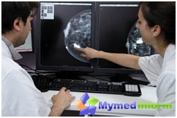
Currently, medicine uses a number of complementary research methods that allow for a short time to put an accurate diagnosis and identify (or reject) those or other pathological changes in lactic glands. But at the same time, only a doctor can make the correct assessment of the results obtained. That is why in identifying during the self-examination of certain changes and the presence of the above-listed symptoms, women should be referred to a mammologist (or oncologist).
In addition, an additional survey may require a surgeon, a gynecologist, an endocrinologist and therapist. After acquaintance with the complaints of the patient, the mammologist's mammologist will appoint an additional study that is most informative in this case.
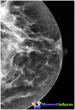
Mammography It occupies a special place among the diagnostic and represents a low and absolutely painless radiographic study of the mammary glands, conducted on a special apparatus - digital mammographer in two projections (for each breast). Currently, digital mammography is considered one of the most informative, affordable and accurate methods for diagnosing the pathology of the mammary glands. Thanks to this study, it is possible to identify small, not even determined when palpation tumor formations in the breast. This method is especially valuable for large volumes of the mammary glands, as well as to detect deeply occurring tumors. But with the preventive purpose, Mammography is recommended between the ages of 38 and older, with the diagnostic - to appoint a physician at any age, starting with 16 years.
Ultrasound of the mammary glands - absolutely harmless to the body of the diagnostic method, in addition, it is painless and widely accessible to women with any state of health and at any age. This method is valuable when conducting a puncture biopsy, as it gives a real-time image, allows you to control the course of the needle to the tumor and evaluate the degree of emptying of the cyst. Also, the ultrasound of the breast vessels with a color doppler mapping is widely used for clarifying diagnostics, which makes it possible to judge the condition of blood flow in a normal and pathologically changed area of the gland, lymph nodes, etc. D. The method is very informative, as it allows you to identify small color objects up to 3 mm diameter. Ultrasound is widely used when examining patients with mammary implants.
Breast dotography - It is a radiographic study in which a special contrast agent is introduced into the breast ducts and a series of pictures in different projections is carried out. By degree, form and contours of filling in ducts, the presence of essentials, extensions or defects of filling are judged on the available (or missing) intra-prototypes (polypa, tumor and t. D.). This type of study is clarifying and carried out in the presence of discharge from the nipple, its deformation by a mammologist.
Penal biopsy, followed by the study of the cytology of the obtained Point - Method conducted by a mammologist, oncologist or surgeon under the control of ultrasound scanning or digital mammography in the presence of unclear nodal formations, tumors and cysts.
These are the main methods of diagnostics, allowing to identify the presence of pathological changes in lactic glands, clarify their structure and character, as well as the prevalence (development stage) of the process. They can be held once (in the presence of indications) or with certain frequency to assess changes during the treatment.
Mammography and ultrasound with prophylactic purposes
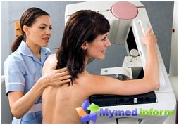
Russian mammologists-oncologists believe that a prophylactic mammography should be carried out by women over 40 years old once every two years, and women of the same age group, but a member of the risk group - once a year. Young women up to 35-40 years old with a prophylactic goal pass the ultrasound of the mammary glands.
And do not forget that self-examination and timely completed additional studies appointed by your doctor will help keep not only health and beauty, but also life. be healthy!


